This is the full version of the article.
This is the second article of a two-part series. Part 1 (https://bit.ly/5411-Insight) covers a snapshot of key historical developments in the Department of Anatomy, National University of Singapore (NUS). This article comprises interviews conducted by Joycelyn Soo (JS) and Helen Cai (HC) with previous and current Heads of Departments (HODs) of the NUS Department of Anatomy – Emeritus Prof Ling Eng Ang (LEA), Prof Bay Boon Huat (BBH), and Prof George Yip (GY) – and an NUS medical alumnus and practising emergency medicine doctor, Dr Darius Pan (DP).
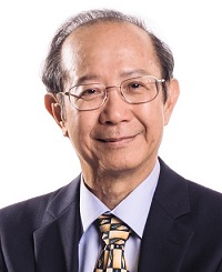 Emeritus Prof Ling Eng Ang attained his Bachelor of Science Degree in Zoology from the National University of Taiwan in 1966, his PhD from the University of Cambridge in 1970 and moved onto a postdoctoral fellowship programme at McGill University. In 1972, Prof Ling started at the NUS Department of Anatomy as a lecturer, teaching and training many batches of doctors and medical students since. During his time as HOD from 1998 to 2008, he made many notable contributions, one of which is developing the Anatomy Museum into one of the department’s best learning resource centres. In addition to his role as an educator, Prof Ling is an accomplished neuroscientist and is known for his research into the biology of microglial – he was the first scientist to demonstrate that microglial cells are derived from monocytes.
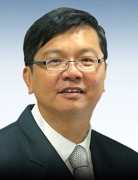 Prof Bay Boon Huat began his journey in education as a senior tutor at the NUS Department of Anatomy in 1989, and he later became the HOD in 2013. During his time teaching at NUS, Prof Bay adopted a student-centred approach. He developed a handbook for pharmacy students and video demonstrations for pre-laboratory sessions. He uses a clinically oriented approach when imparting knowledge about anatomy and emphasises on building basic foundational concepts to medical students. As a scientist, Prof Bay’s research focuses on the biology of cancer, specifically the utility of biological markers of malignancy. As a highly recognised scientist, he has been invited to author book chapters such as “Embryology and surgical anatomy: Thyroid and parathyroid glands” and to present his work at international microscopy conferences.
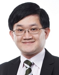 Prof George Yip is the current HOD of the NUS Department of Anatomy. He has been greatly involved in the education of students, being the coordinator of the Undergraduate Research Opportunities Programme in Science and Honours Project in Life Sciences (LSM4199), and his commitment to education excellence has been recognised by the NUS Annual Teaching Excellence Award and the Yong Loo Lin School of Medicine Faculty Teaching Excellence Award. His research mainly lies in the roles of glycosaminoglycans and proteoglycans in cancers and wound healing. In the area of academia, he was awarded the International Fellowship award by A*STAR in 2002–2003 and the Japan Society for the Promotion of Science (JSPS) Fellowships in 2005 and 2011, won the Clinician Scientist Award twice in 2012 and 2019, and has served on the Editorial Board of many journals.
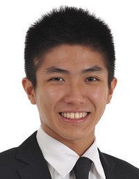 Dr Darius Pan graduated with honours from NUS in 2016 and is an emergency medicine doctor practising at NUH. He was appointed adjunct lecturer in Anatomy under the Clinician Educator Scheme in 2022.
JS, HC: Hello Prof Ling, Prof Bay, Prof Yip and Dr Pan. Very nice to meet you and thank you all for your time today. We hope to commemorate 100 years of NUS Anatomy with this article, sharing the insights and reflections of our educators and former students at the NUS Department of Anatomy.
To start off with a question to our professors, what did you enjoy most about teaching at NUS Anatomy?
LEA: I am not a medically trained person, having been trained as a histologist and neuroscientist. After getting into the course of anatomy however, I grew to like it and could integrate the various disciplines of anatomy, including gross anatomy (neuroanatomy in particular), histology and embryology. I found that once the knowledge is integrated across the various disciplines, one comes to like it and the students seem to be able to appreciate it better. I initially spent most of my time doing histology lectures but of course later I did gross anatomy. Over the years, I found I could appreciate the subject better and thanks to COVID-19 when I was forced to stay home, I spent a lot of time on YouTube learning to better integrate anatomy and physiology.
July 2022 marked 50 years since I joined the department. We used to only teach medical, dental and biology students back in the 1970s but now we also teach the other healthcare professional students. For me, the best part is when the students appreciate that we have taught them. Better yet is my attending doctors being appreciative when I go for my medical check-ups. I remember once when I emailed my ophthalmologist, who has been taking care of me for the long term, to thank him, and his reply was, “Thank you for teaching us so we are now able to help you”. In fact, I very much enjoy teaching. That’s why I am still here at NUS. If I could teach as a hobby, I would feel blessed.
Dissection and the Silent Mentors
JS, HC: Thank you Prof Ling. We are most appreciative that our tutors in Anatomy are committed to education, motivating us with their passion for anatomy. We know that the department has been constantly improving its methods of education and have started new programmes over the years. One of these efforts is the Body Donation programme. How did that get started?
LEA: Let me introduce a bit of our history of using cadavers. We started off with using cadavers in the 1980s as we had a supply of cadavers from the Health Sciences Authority. Most were unclaimed bodies but the number of bodies started dwindling over the years. We could not afford having every student perform dissections, and another concern was fitting over 180 students in the dissection hall. Around the year 2003, we thought that one way to preserve the number of the cadavers would be to use prosections. We engaged staff from Kunming, China to prepare prosections for students’ learning, which was called “exploratory learning” back in my time. However, the number of available cadavers was still declining until we started the Body Donation programme.
Additionally, not every student is keen to dissect. We used to have seven or eight students to one cadaver – which was quite a luxury. However, maybe two or three of them may have never actually performed a dissection and yet, they were still able to pass the examination. That would be a waste of the precious resources.
Currently, Year 1 students look at prosections and learn the anatomy. During the summer break, dissection is available as an elective for Year 1 students. Those who are keen to dissect can opt to come back for it, and the attendance has been quite good. In the old days, we used to patrol to see whether students dissected correctly and assist them. Since students are willing to come back and learn, and we have enough Silent Mentors from the Body Donation programme, this arrangement is quite ideal.
If you ask me, dissection is the way to learn anatomy – you learn by looking at the person. In Chinese, there is a popular saying, “百闻不如一见”, and more recently, “百见不如动手”, which means seeing once is better than hearing a hundred times, and getting into the action and doing is better than just seeing. On top of that, when new lecturers go around during dissection, they are also learning while they teach, and learning becomes mutual.
BBH: Prof Ling has actually revolutionised this whole aspect. We were very fortunate that he had the foresight in 2003 to shift to prosection. If we did not have all these prosected specimens during the COVID-19 pandemic, everything would have come to a standstill due to cadaver shortages.
The Silent Mentor programme was adapted from Tzu Chi University as we found the concept particularly good and applicable to Singapore’s context. We wanted our students to appreciate their Silent Mentors when undergoing dissection sessions, and to engage in events post-dissections to show their appreciation.
DP: Back when I was a first-year medical student in 2011, the Silent Mentor programme was still in the developmental stages. Ten years later, as I return to the department to teach, many things have changed. For example, the faces and names of the Silent Mentors are on a mural outside the Anatomy Hall, and beside the respective cadavers as well. That way, we put a name and age to the Silent Mentors during sessions, so that is very meaningful especially for people who want to donate their bodies under the Medical (Therapy, Education Research) Act. Essentially, the cadaver is like our first patient. Through the Silent Mentor programme, we learn to show respect and appreciation for our patients, and this sets the tone which carries through to the next phase of our career from students to doctors.
JS, HC: Yes, we have heard that dissections were considered the norm in teaching anatomy back in the day, as well as animal laboratories. Could you share more with us on how that was like?
LEA: We used to have some rats and monkeys at the former Sepoy Lines Campus. When we moved to the current Kent Ridge Campus in 1983, we kept the primates, and I did some research with the animals. We used the perfusion method to put the animals to death.
As anatomists, our research in the past focused on experimental morphology. Gradually, it has been shifting to biochemical, pharmacological, and biomolecular research, where people are increasingly doing tissue culture and using samples from the hospital. We still do some research with Tan Tock Seng Hospital, where the radiologist chief was also our student.
Even till today, I would like to emphasise the old saying that anatomy is the geography to medicine. I often tell the people around me this: when you hold someone, touch someone, look at someone, that is all anatomy. The anatomy discipline, including histology and neurology, is extremely important not only for medical practitioners, pharmacists and dentists, but is also very relevant for any graduate student who wants to do research.
New approaches to anatomy
JS, HC: Yes, we absolutely agree that we see anatomy every day and can learn from it just by observing. With the rapid expansion of medical knowledge in the current era and with the changing responsibilities of doctors, how has the teaching of anatomy at NUS changed?
BBH: I think nowadays the approach is more towards practical application and knowledge. There is no point in learning all the individual branches for the arteries, but it is good to know the functions and significance of the blood supply. We are now more aligned towards clinical anatomy. You will find that our tutorial objectives are based on clinical scenarios and how you would apply what you have learnt. That is what I think students would benefit from the most when they go to the wards. They can relate the anatomy that they have learnt to what they are seeing; it does not make sense for them to just learn the anatomical structures by rote memory. This is also what medical schools around the world are doing.
LEA: When I first joined in the 1970s, the total number of anatomy contact hours were over 700 hours. It is now being gradually reduced to about 200 hours. To me, a great outcome from our many medical curriculum reviews is that we trimmed away a lot of the unnecessary details. If you look at our tutorial objectives now, you will see that they are much more functional, focusing on applied anatomy with clinical relevance. If not, you may end up memorising all the branches of the brachial plexus without knowing what their relevance is. For example, we have done away with all the cutaneous branches, and that knowledge is more than enough for most practising GPs. If the students want to become specialists, they can go into more detail later. We have also simplified the curriculum – retaining its essence while making the content more clinically relevant.
We were lucky to have Prof Rajendran K, a surgeon, help us a lot in refining the tutorial objectives. Another benefit of this change is that we are now able to better integrate anatomy with the other disciplines.
Nostalgic memories
JS, HC: We have heard about how life is like for educators at NUS Anatomy and were wondering if Prof Yip could share with us how it was like as a former student and now as an educator. Do you have any fond memories during your time as a student?
GY: Well, I do have this interesting story. Most of the lecturers back then used blackboards and overhead projectors. The Omni-Theatre at the Science Centre and planetarium started operation not long ago then. They used 35 mm slide projectors at the planetarium to let visitors get a sense of the changes in the stars by turning on the projectors alternately. It felt like the fade transition that you get from your PowerPoints these days.
So, for anatomy back in those times, Prof Rajendran would bring two 35 mm projectors to class. He was very artistic and used plasticine to make models for both gross anatomy and embryology teaching. By making use of both projectors, he would demonstrate the transition between layers and reveal underlying structures after removing different layers. Using the same approach, he could also show changes in different parts of the embryo, such as how the midgut herniates out, undergoes rotation and goes back in again during gut development.
LEA: The prototype for animated transitions was used by Prof Voon and Prof Rajendran. They used many layers of transparency and drawings and flipped through them just like it was in 3D. They used this to show gut rotation and heart formation, and used plasticine models to produce different parts of the body including knee joints. So yes, I would say the department has some really clever teachers.
JS, HC: Dr Pan, could you share with us your fondest memory at NUS Anatomy?
DP: Anatomy was one of the first subjects we were exposed to in pre-clinical years and one of the core topics in our first year of medical school. Entering first year, most of us were wide-eyed and ready to learn, and anatomy is an especially good fit because it introduced us to the human body – its structures and how it functions. I still remember the Latin names that we had to learn; those left a very deep impression. And moving through our first year, we got to see the cadaveric specimens and microscopic specimens at the laboratories, which gave us insight into the “very big” and the “very small”.
I remember when I was in Year 1, our seniors would tell us about the “Papa”, “Mama” and “Baby” versions of the medical textbooks which differ by their thickness. As anatomy textbooks were very thick, I bought the “Mama” version. For convenience, I would cut the textbook into chapters and bring these to read on the train and into the lecture halls.
My fondest memory in anatomy is of the time I spent with my tutor, Dr Satish LR. We struck a very deep friendship over the past decade. He is the sort of tutor who encourages you to never stop learning. When we asked him questions which he did not have the answer to, he would take it as a learning point for himself and go on to investigate and return with answers during the next tutorial. I am thus inspired by him to never stop learning and to take every opportunity further my knowledge.
JS, HC: Agreed, Dr Pan. Anatomy is the first thing we get to know in medicine, and it sticks with us throughout our careers many years after we graduate. Speaking of this, how has the Anatomy department still been relevant in your training as a doctor?
DP: I remember it was during my medical officer year when I met Dr Satish in the wards while he was taking students on their patient-based learning. That was also how I got involved in teaching, taking the pre-clinical students to see patients and, from there, learn clinical anatomy. The emergency department (ED) is where we get the first contact of all patients and we can look at the X-rays, ECGs and other imaging of our patient and see how we can clinically correlate this for teaching during patient-based learning. From there, I also had the opportunity to teach at the Silent Mentor dissection section such as anatomy and airway management. Some procedures applicable at the ED are surgical airway, inserting the endotracheal tube during intubation and insertion of chest drains; these opportunities are very precious are hard to come by even for ED residents, but during cadaveric courses, we are able to learn and conduct these procedures at the level of M2.
To another 100 years!
JS, HC: Two final questions before we end the interview. What do you hope for the Anatomy department to achieve in the coming years, and do you have any well wishes or congratulatory words for the department?
DP: The evolving focus of anatomy will be on more intentional clinical application and its foundational role in clinical practice. Studying medicine is like reading a novel and anatomy is like the first few chapters of it; sometimes in the first few chapters, we have no idea what is going on, and may find it boring. What we hope to do is to drop hints of the later years’ ongoings in Year 1 so that students will be able to understand the final application of anatomy in patient care. In future years, we will also move towards a more systems-based approach in teaching anatomy.
The NUS Department of Anatomy has been a long-standing institution and its heritage can be traced back to the pre-war era. I congratulate the department on the past 100 years, and hope that the department can continue to impart important knowledge to future generations of doctors and students.
GY: The department has come a long way since it was established. The NUS Anatomy department has been recognised as one of the leading institutions in Asia. Moving forward, we would like to build on this and become even better not just in Asia, but also in the global arena.
LEA: Our primary aim is really to teach our students well. As one of our vice-chancellors used to say, “teachers exist because of students”.
JS, HC: Thank you all very much for your time today, and for sharing with our readers. We congratulate NUS Department of Anatomy on their centennial celebration!
